Have you seen this type of image anywhere, like in any hospital or at somebody's home? You must be wondering: What is this, what type of images are these, and how are they formed?
Have a seat, because I am going to explain everything here.
What is CT scan?
A CT scan, or, in full form, a computed tomography scan, is the combination of X-ray images taken from different angles to create detailed cross-sectional images of the inside of the body. It's commonly used to diagnose various medical conditions, including injuries, tumors, and internal bleeding. Basically, it's an X-ray, but in video mode. Like when you are taking photos and videos. Videos are collections of photos at higher speeds; likewise, CT-Scan is the collection of X-ray images at higher speeds.
How these Images are formed ?
The image you were seeing is from the above machine, where you get this type of image. It is called a CT scanner or Computed Tomography scanner. It basically uses the same principle as X-ray imaging, but it is more advanced, useful, and accurate. If you want to know about X-ray imaging, then
What is X-ray imaging?
Here you can see a big, round part called the rotating X-ray source (tube) or Gantry. From where the X-ray comes. It is rotatable. And you can also see a bed-like platform that is motorized and movable to the front and back. So when we switch on the machine, the movable platform goes in front, and the rotating part captures the body's images from all directions. And we can capture images of the internal structures of the body. Thus, a CT scan is nothing but a 3D X-ray image of a part of the body, or you can scan the whole body through it.
Images are taken from various angles. Gantry has an x-ray tube and detector. The table goes in, and an x-ray tube rotates around the table. Most commonly used CT scan is 3rd generation CT scan.
The distance the table moves for every rotation of the gantry is known as pitch. Pitch is inversely proportional to the quality of the image and the dose of radiation.
So, How can we read it?
First, I am going to give you some information.
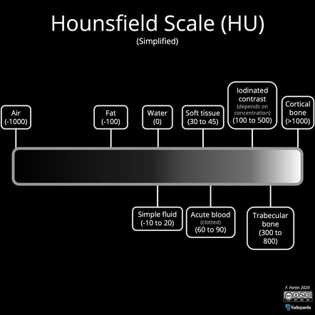
The CT scan was invented by Godfrey Hounsfield, so based on his name, we have given the unit of attenuation (stopping) of the X-rays, called Hounsfield units (HU).
Its the measurement of radio density of the substance.
We have to make a reference material, if we are calculating density of any substance, So we take distilled water a reference.
Thus, the HU value of distilled water is 0.
Positive and negative HU values are given based on the density compared to water. Bones have a high positive HU value (+1000), and air has a high negative HU value (-1000).
Soft tissue (+40) and metals have a higher HU value than bones (maximum white),
We compare fat with oil, and oil is less dense than water, so fat has a negative value (-10 to -100).
In a CT scan, different tissues absorb X-rays differently.
- Air appears black.
- Fat appears dark gray.
- Water and soft tissues appear light gray.
- Bone and calcifications appear white.
- Metal and other dense materials are very bright whit
Black is considered hypodense and white is considered hyperdense.
In this picture, you can see a white borderline, which is the skull. See the whitness or opacity of the bone. The inner part is soft tissue, which is gray in color. And black lines are fat tissue or CSF flowing through the ventricles.
Now, What are the types of CT scan we can see:
There are several types of CT scans, each examine to specific areas of the body or medical conditions. Some common types include:
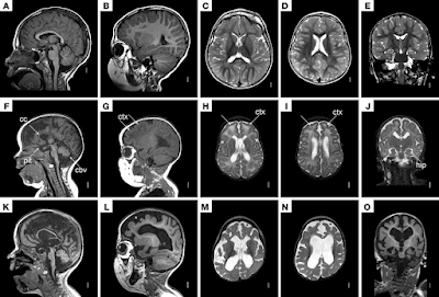
1. Head CT (Cranial CT):
As the name suggest, It is used to examine the brain and skull for injuries, bleeding, tumors, infections, and other abnormalities.
2. Chest CT (Thoracic CT):
It provides detailed images of the chest area, including the heart, lungs, and surrounding structures. It can help diagnose conditions such as lung cancer, pulmonary embolism, and pneumonia.
3. Abdominal CT:
It focuses on imaging the abdominal organs such as the liver, kidneys, pancreas, spleen, and intestines. It is used to diagnose conditions like abdominal pain, kidney stones, appendicitis, and liver disease.
4. Pelvic CT:
It is used to examine the pelvic region, including the reproductive organs, bladder, and rectum. It can help diagnose conditions such as ovarian cysts, uterine fibroids, and prostate cancer.
5. CT Angiography (CTA):
It is specialized CT scan that focuses on imaging blood vessels to detect blockages, aneurysms, or other vascular abnormalities. It is commonly used to evaluate the arteries in the brain, neck, heart, and extremities.
6. Virtual Colonoscopy (CT Colonography):
It is a non-invasive alternative to traditional colonoscopy for screening and detecting colon polyps or cancer. It uses CT imaging to create detailed images of the colon and rectum.
7. Cardiac CT:
It is specifically designed to assess the heart and coronary arteries for conditions such as coronary artery disease, heart valve abnormalities, and congenital heart defects.
 |
| Gastric CT |
 |
| Heart CT |
 |
| Heart CT |
 |
| Lung CT |
 |
| Brain CT |
These are just a few examples of the types of CT scans available. The specific type of CT scan recommended will depend on the patient's symptoms, medical history, and the area of the body being examined.
There can be an another type, called Contrast enhanced CT(CECT).
It is unique and important because in this we use contrast agents, mostly iodinated contrast,which shows a white color on the CT scan. It helps identify lesions, inflammation, or infection pathology in any organ. It gets deposited and shows a white color on the report.
Now if we use X-Rays in this also,
How and why it is different from X-Ray imaging?
First, CT scan shows 3D view of the part, in case of X ray imaging, we can see in 2D.
Second, It shows clear picture of the part compare to X-Rays.
Lets discuss with images,
 |
The first image shows an X-ray of the chest. The second image is a CT scan.
If we had not done the CT scan, then the doctor may have thought it was a tumor. |
 |
Its also an X-Ray of Chest and CT scan of chest
Can you see the difference between them? |
But, till date, we have used X-ray machines because they are cheap and they are an initial investigation for the doctors.
Thus, this end my explanation. If you have any doubt, please reach me out on my contact us page.
If you want to know about any other radiological findings
What is X-Ray?
What is Sonography?
Thank you


.jpeg)
.jpeg)


.jpeg)
.jpeg)

0 Comments