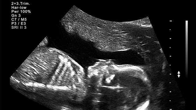
You must have seen this type of image in someone's common room or bedroom as a frame. Can you identify it? It's an ultrasonography of the fetus, maybe about 20 weeks. They keep it as a momento. Because it's the first image of their child.
But ultrasonography is not only done during pregnancy; it is also used to detect various diseases and complications. Like kidney stones, gall bladder caliculi, detection of cysts, etc., we will discuss them in detail.
What is Sonography or Ultrasonography?
Ultrasonography/ Sonography/ultrasound, is a medical imaging technique that uses high-frequency sound waves to create images of internal body structures. It's commonly used for diagnostic purposes, such as examining organs, tissues, and blood vessels, as well as monitoring pregnancies.
It contains many technical devices to examine the patient, it includes
Probe:It's the handheld device that the sonographer (technician) moves over the body to capture images.
Console: This is the main unit of the ultrasound machine where the sonographer controls the settings, captures images, and reviews the results.
Display screen: The ultrasound images are displayed in real-time on a monitor, allowing the sonographer to observe and interpret the images during the examination.
Control panel: This includes buttons and knobs that allow the sonographer to adjust settings such as depth, focus, and frequency of the ultrasound waves.
The sonographer also uses gel on the surface, which is helpful for two purposes: first, it acts as a lubricant, and second, it acts as an interface for the conduction of sound. Air is a bad conductor of sound, so we need a water-based gel to remove air in between the probe and the body.
How does Ultrasonography works?
First, answer this: Have you been to any large auditorium, and when you shout your name, it repeats back? The phenomenon happening here is called echo. You must have studied in your school days, i.e., the reflection of original sound bounced back from any surface and returning to the listener's ear cause echo.
The exact same phenomenon happens in the case of ultrasonography.
It works on principle known as Pulse echo or Piezoelectric effect.It's a phenomenon where certain materials generate an electric charge in response to mechanical stress or pressure. This effect is due to the rearrangement of the crystal structure of the material when it is subjected to mechanical force, resulting in the separation of positive and negative charges within the material. Piezoelectric materials are commonly used in various applications, such as sensors, actuators, and transducers.
So when the Pulse of sounds are sent to the patient's body.These sounds are reflected from various tissues interfaces.The reflected sound waves are converted in to electrical signals and we can see the internal structure on the monitor.
The Ultrasonography probe converts the electrical signals to sound waves and sound waves to electric signals by piezoelectric effect.The probe surface has the piezoelectric crystal made of leadzirconium titanate.
If you notice, I am constantly saying sound wave. If it's a sound wave, then how can't we listen to it? The answer is simple: humans generally can hear sound waves up to 20 Hz to 20 kHz. And we use approx. 1-2 MHz in ultrasonography.
Now How can we read a Sonograph?
I am mentioning some term, which we hear in Sonography investigation:
Echogenicity:
It is the term used to describe the abililiy of different structure or tissue to reflect sound wave.
For example:
1.Hypoechoic: Tissues that appear darker or less bright than surrounding tissues on an ultrasound image are described as hypoechoic. This indicates that the tissue reflects fewer ultrasound waves.
2.Hyperechoic: Tissues that appear brighter or more reflective than surrounding tissues are described as hyperechoic. This suggests that the tissue reflects more ultrasound waves.
3.Isoechoic: Tissues that have the same level of echogenicity as surrounding tissues are described as isoechoic. This means they have similar reflectivity to ultrasound waves.
4.Anechoic: Structures or regions that appear black on an ultrasound image are described as anechoic. This indicates that they do not reflect any ultrasound waves and appear as
fluid-filled areas.
Modes of Ultrasonography:
A-Mode(Amplitude Mode):
It is one of the earliest and simplest forms of ultrasound imaging. In A-mode, ultrasound waves are sent into the body, and the returning echoes are plotted on a graph showing the depth of the tissue or structure (y-axis) against the amplitude of the echoes (x-axis).
A-mode ultrasound is primarily used for measuring distances and dimensions within the body, such as the thickness of tissues or the depth of organs.
It's commonly used in ophthalmology for measuring the length of the eye and in certain medical procedures such as detecting foreign bodies or guiding needle placements during biopsies or injections.
B-mode ultrasound:
It also known as brightness mode ultrasound, as the name suggest, the waves are sent into the body, and the returning echoes are used to generate a two-dimensional grayscale image of the scanned area in real-time.
In the resulting image, different shades of gray represent variations in tissue density, with
brighter areas indicating higher reflectivity or density and
darker areas indicating lower reflectivity or density.
M-mode ultrasound:
It is also known as motion mode ultrasound, is a specialized mode of ultrasound imaging that displays motion over time along a single ultrasound beam. In M-mode, ultrasound waves are sent into the body, and the returning echoes are used to create a one-dimensional image where movement is displayed along one axis, typically time, while depth is displayed along the other axis.
M-mode ultrasound is particularly useful for assessing the movement and function of specific structures within the body, such as the heart, valves, and blood vessels.
Doppler mode ultrasound:
It is a specialized mode of ultrasound imaging used to evaluate blood flow within blood vessels. It utilizes the Doppler effect, which involves changes in the frequency of sound waves reflected from moving objects, such as red blood cells.
Here colors are assigned to different flow velocities, allowing for the visualization of blood flow patterns and directionality within the vessels. This mode is particularly useful for assessing overall blood flow and identifying abnormalities such as stenosis (narrowing) or occlusion (blockage) within blood vessels.
 |
In this image, you can see blood flow. If the blood flows in the direction of the probe, it shows red, and if it flows opposite to the probe, it shows blue. |
Posterior acoustic shadowing:
Lets understand it with an Example:
Here, in this image, you can see that the patient is suffering from a gall bladder stone (caliculi), as the sound wave reaching the stone is completely reflected. So no sound is reaching the area behind the stone, thus there is no echo, no image, and the area appears black, indicating a shadow.
This phenomenon called posterior acoustic shadowing.
E.g
- Renal calculi- calcified.It shows clear shadowing.
- Air-bad conductor, so it reflects all the sound.It shows dirty shadowing
- Bones-calcified,so gives shadow. It shows clear shadowing.
Posterior acoustic Enhancement:
Let's take another patient. She has a cyst in her breast; this is how we identify it.
In this case, it shows white areas posterior to the structures. Which is seen with fluid-filled lesions called cysts. Mainly simple cysts.
We know fluids are good conductors of sound.
So there is no reflection of sound from the cysts. No echo-anechoic.
As the cyst is filled with fluid, it transmits all the sound waves through it. So the area behind the cyst gets more sound waves, thus brightening that space.
This phenomenon is called posterior acoustic enhancement.
Now we have read Echogenicity, Modes, shadowing and enhancement.
If you combine all this and see a real ultrasound report, i am sure you can read it, with some practice.
Ultrasonography has many advantages, like:
- It uses sound waves therefore, there is no radiation,
- It is a preferred investigation in pregnancy.
- It is cost effective.
- It is easily available.
- It can be Portable.
Here are some images of Sonography
 |
| Ultrasound during early pregnancy |
 |
| Endometrium in various phases of menstrual cycle |
If you want to know about radiation investigation like X-rays and CT scan;
Thus, This is a basic explanation about Ultrasonography. Hope you understand.
Thank you


.jpg)
.jpg)
.jpg)


.jpeg)
.jpeg)


.jpeg)
.jpeg)

0 Comments