The above image, which you are seeing, is an MRI of a normal 3-year-old boy. If you have any background in biology or medicine, you would be excited because you can see the brain cortex, eyeball, ventricles, medulla oblangata, etc. MRI is kind of a blessing to medical science because, without it, it would be very difficult to identify and diagnose an internal disease. Think of a brain tumor, where there is an abnormal growth of tissues inside the brain. How can a doctor say the location of the tumor and how it is affecting different parts of the brain?
MRI is the best diagnostic method to identify any abnormality in soft tissue. Of course, there is a CT scan, ultrasound and X-rays, but it generally doesn't show an exact or high-resolution image of the abnormality.
What is MRI?

MRI or Magnetic Resonance Imaging, is a medical imaging technique used to visualize internal structures of the body in detail. It uses a powerful magnetic field, radio waves, and computer technology to generate detailed images of the internal structures of the body. It provides high-resolution images of soft tissues, organs, and bones, allowing healthcare professionals to diagnose and monitor a wide range of medical conditions. MRI is especially useful for imaging the brain, spinal cord, joints, and muscles, and it does not involve ionizing radiation,like CT scan and X-rays, making it a safe option for most patients.
It mainly composed of :
Magnet:
It is the main component of the MRI machine, which generates a strong and uniform magnetic field. This magnet is typically a superconducting magnet cooled by liquid helium to maintain its superconducting state.
Gradient Coils:
Gradient coils are additional coils within the MRI machine that produce varying magnetic fields in different spatial directions. These gradients are used to encode spatial information into the MRI signals, allowing for the creation of detailed images.
Radiofrequency (RF) Coils:
RF coils are used to transmit radiofrequency pulses into the patient's body and to receive the resulting MRI signals. These coils can be placed around the body part being imaged or within specific areas of the MRI machine.
 |
| RF coil around patient's head |
How does it Work?
Have you heard of Magnetic Resonance concept. It refers to the behavior of atomic nuclei, such as hydrogen nuclei (protons), in a magnetic field. When placed in a magnetic field, these nuclei align themselves with the field, creating a net magnetization in the direction of the field.
When an external radiofrequency (RF) pulse is applied, it can cause the nuclei to absorb energy and temporarily shift their alignment. After the RF pulse is turned off, the nuclei release the absorbed energy and return to their original alignment. This process of absorbing and releasing energy is called resonance.
Similarly, in the case of MRI, firstly, a radiofrequency coil( RF coil) is placed around the patient's. It produces a radiofrequency pulse into the patient's body. This patient is then sent inside the MRI machine, which contains a large magnet that produces a powerful magnetic field.
When a patient enters the MRI machine, their body's hydrogen atoms (protons), which are abundant in water and fat molecules, align with the strong magnetic field generated by the MRI magnet.
Radiofrequency (RF) coils emit short bursts of radio waves at specific frequencies. These RF pulses are directed into the body, causing the aligned protons to temporarily absorb energy and flip their alignment.
After the RF pulse is turned off, the protons gradually release the absorbed energy and return to their original alignment with the magnetic field. During this process, they emit radiofrequency signals, which are detected by the RF coils in the MRI machine.
The emitted signals are received by the RF coils and converted into electrical signals. These signals contain information about the density and distribution of protons in different tissues within the body. Thus,creating a detailed image of internal organs on monitor.
The magnetic strength of MRI machines is indicated in TESLA (T). Thats why in a diagnostic center,you must have heard 1.5 TMRI, 3 TMRI, etc.
Types of MRI:
There are several types of MRI,each serving different purpose and providing specific information about the body's internal structures.But there are some common types like:
T1-weighted MRI:
This type of MRI scan provides detailed anatomical images with excellent contrast between different types of tissues. It is useful for visualizing anatomy and detecting abnormalities like tumors, hemorrhages, and structural changes.
T2-weighted MRI:
T2-weighted MRI scans highlight tissues with high water content, such as cerebrospinal fluid (CSF), edema, and inflammation. They are often used to diagnose conditions like brain lesions, spinal cord abnormalities, and joint injuries.
FLAIR MRI :
It is commonly used in neuroimaging to improve the visualization of brain lesions, particularly those located adjacent to CSF-filled spaces. It can enhance the detection of abnormalities such as multiple sclerosis plaques, brain tumors, and inflammatory lesions by suppressing the signal from surrounding CSF, thereby increasing lesion conspicuity.
Diffusion-weighted MRI:
Diffusion-weighted MRI measures the random motion of water molecules in tissues. It is sensitive to changes in tissue microstructure and is particularly useful for detecting acute stroke, tumors, and white matter abnormalities.
Functional MRI (fMRI):
Functional MRI is a specialized technique used to measure brain activity by detecting changes in blood flow and oxygenation. It is commonly used in neuroscience research to study cognitive functions, language processing, and neurological disorders like epilepsy and Alzheimer's disease.
Now, I have explained the basics of MRI: what is MRI?, how MRI works?, and its types.
So, how is it different from a CT scan?
.jpg)
The first image in the photo is a CT scan, while the second is an MRI. Can you differentiate it?
Firstly, see the resolution. In the case of an MRI, we can see the soft tissue, like the liver, heart, spleen, etc., more clearly, so it is preferred in cases of soft tissue abnormalities.
Second, a CT scan uses radiation like X-rays, which can be dangerous, but in the case of an MRI, it uses a magnetic field, which is safer.
Third,MRI offers functional imaging techniques such as functional MRI (fMRI), diffusion MRI (dMRI), and magnetic resonance spectroscopy (MRS), which provide information about brain activity, tissue microstructure, and metabolic processes. These techniques are not available with CT scans and are valuable for studying neurological disorders, tumors, and other conditions.
Fourth,MRI allows imaging in multiple planes (axial, sagittal, and coronal) without repositioning the patient. This flexibility is advantageous for visualizing anatomical structures from different perspectives and can improve diagnostic accuracy.
So, why don't we use MRI for often, there are some contraindication, thats why.
Like,
- Patients with cardiac pacemakers
- Cochlear implant.
- Aneurysmal clips.
- If the patient has Claustrophobia, MRI machine is like a tunnel.
- Recent metallic implants and prosthetics.
- Tattoos on the body, a relative contraindication - Generate a lot of heat
- Dental implants generate artefacts, when MRI brain is done.They degrade the image quality
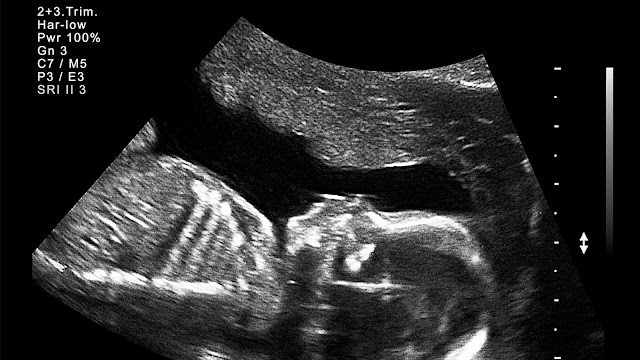
Here are some images of MRI of normal person
 |
| Pons |
 |
| Medulla |
 |
| Anatomy of Midbrain |
 |
| Mid sagittal brain |
 |
| Female reproductive System |
 |
| Uterus |
If you also want to know about CT scan, X-ray imaging and Ultrasound:
Thank you


.jpg)


.jpeg)
.jpeg)


.jpeg)
.jpeg)

0 Comments